Leg pain does not automatically mean that the arteries supplying your legs are narrowed. The timing, location and triggers of the pain—along with changes in skin colour, temperature, pulses and wounds—help distinguish arterial disease from nerve, joint, muscle and vein problems. By the end, you will know which symptoms need urgent care, which tests clinicians use and how to prepare for an evaluation.
Key takeaways
- Walking pain that eases with rest fits intermittent claudication.
- Compare PAD symptoms with restless legs, venous disease and sciatica.
- Seek prompt assessment for rest pain, foot wounds or colour changes.
- An ankle-brachial index often starts the evaluation for suspected PAD.
Does your leg pain follow the pattern of intermittent claudication?
The classic pattern of intermittent claudication is predictable muscle discomfort that starts during walking and eases after you stop. It feels like aching, cramping, tightness or heaviness, rather than pain that appears randomly while you sit or lie down.
The location can help identify the pattern:
- Calf pain is the most common presentation, especially after walking a repeatable distance or climbing stairs.
- Thigh pain suggests reduced blood flow higher in the leg.
- Buttock pain can indicate disease in the pelvic or iliac arteries.
The pain should improve within about 10 minutes of rest, then return when you resume similar exertion. A fixed walking distance is not required, but the relationship with activity and rest should be fairly consistent.
Pain that remains constant, shifts unpredictably, or has no connection with exertion points away from classic intermittent claudication, although it does not rule out peripheral arterial disease.
PAD does not always cause pain. Fatigue, heaviness or a steadily shrinking walking range can be the only clues, particularly if you have diabetes, nerve damage or limited mobility. A normal resting ankle-brachial index also does not exclude PAD when exertional symptoms persist; ask about exercise ABI testing.
How PAD symptoms differ from restless legs, vein problems and sciatica
Pain that follows a nerve, joint, muscle injury or prolonged standing points away from poor arterial circulation. PAD more often causes reproducible calf, thigh or buttock aching, cramping or heaviness during walking; stopping relieves it within about 10 minutes.
| Cause | Clues that point away from PAD |
|---|---|
| Restless legs | An urge to move, crawling or aching sensations while sitting or lying down, and symptoms that worsen in the evening or at night. Movement brings relief. |
| Varicose veins | Enlarged, twisted surface veins with aching or throbbing after standing. Elevating the legs helps. |
| Chronic venous insufficiency | Swelling around the ankles, heaviness, itching, brown skin changes or ulcers near the inner ankle. Symptoms worsen through the day. |
| Sciatica | Burning, electric or shooting pain from the lower back or buttock down the back of the leg, sometimes with tingling or weakness. Coughing or bending can aggravate it. |
| Arthritis | Pain centred in a hip, knee or ankle joint, with stiffness, reduced range of motion and pain during joint movement. |
| Muscle injury | Localised tenderness after lifting, exercise or a sudden movement, often with bruising or pain when the muscle is stretched or contracted. |
These patterns can overlap. Leg swelling favours venous disease, medication effects, lymphedema or systemic illness, but mixed arterial and venous disease also occurs. Do not start strong compression stockings blindly if PAD is possible; ABI or toe-pressure testing helps determine whether compression is safe.
Which warning signs and risk factors make leg pain more concerning?
Sudden severe leg pain with a cold limb, pale or blue skin, absent pulses, numbness or weakness is an emergency. Call emergency services immediately; acute limb ischemia can permanently damage tissue within hours.
| Warning sign | What it suggests | Action |
|---|---|---|
| Pain at rest, a nonhealing foot wound, or gangrene lasting or expected to last at least two weeks | Chronic limb-threatening ischemia | Arrange prompt vascular assessment |
| Black toes, pale toes, a sharply defined ulcer, or a cold foot | Severely reduced arterial flow | Seek urgent medical evaluation, especially with worsening pain |
| A blister, drainage, swelling with redness, or infection in a person with diabetes or neuropathy | Delayed healing and possible tissue infection | Contact a clinician promptly; do not wait for severe pain |
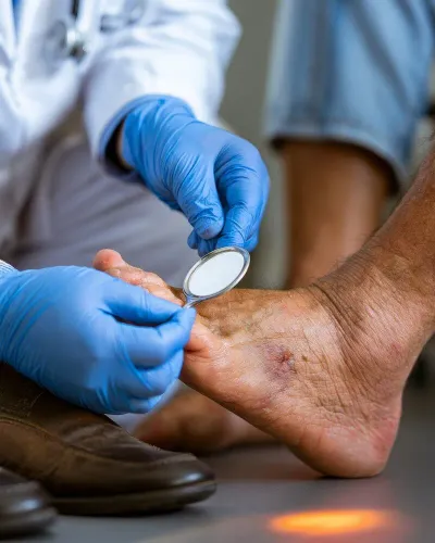
Check your feet every day if you have diabetes or reduced sensation. Protect them from barefoot walking and tight shoes, because neuropathy can hide an injury while poor blood flow delays healing.
Suspicion of peripheral arterial disease rises with these factors:
- Current or previous smoking
- Diabetes
- High blood pressure
- High cholesterol
- Chronic kidney disease
- Older age
- Previous heart attack, stroke, or other cardiovascular disease
Leg swelling alone points more often to vein disease, lymphedema, medication effects, or systemic illness than uncomplicated arterial disease. A cool foot, weak pulses, exertional symptoms, or a toe ulcer deserves vascular assessment, even when swelling is also present.
Which checks confirm or challenge a PAD suspicion?
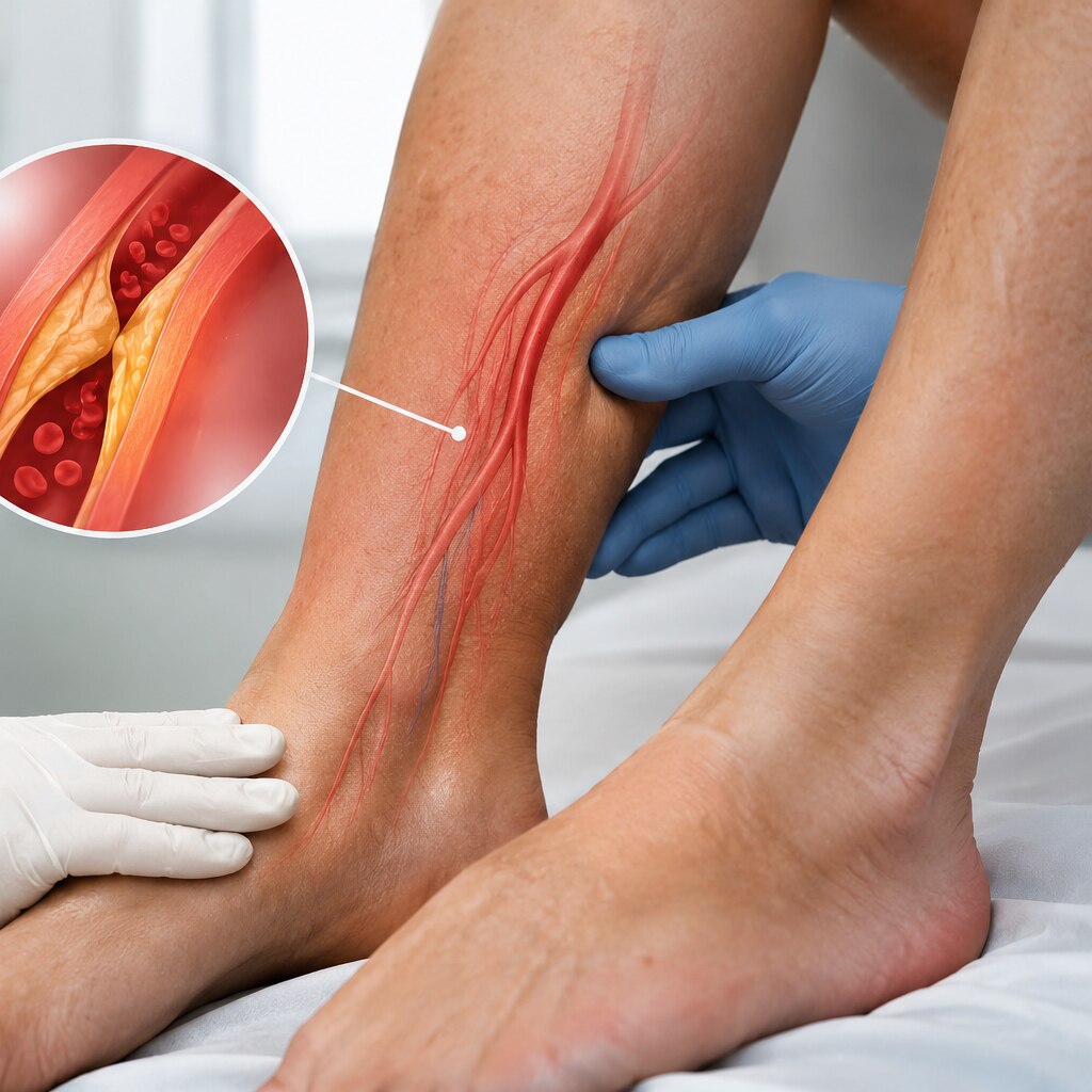
A clinician will compare pulses in both legs, inspect colour and wounds, and check skin temperature and capillary refill. A weak or absent foot pulse, cold skin, delayed refill, or a nonhealing sore strengthens the suspicion but cannot locate the blockage by itself.
For an ankle-brachial index (ABI), avoid walking, smoking and vigorous exercise immediately beforehand. Rest flat and still for 5–10 minutes, wear clothing that leaves both ankles and arms accessible, and take prescribed medicines unless the clinic tells you otherwise. Tell the clinician if you have diabetes, chronic kidney disease or known arterial calcification.
| Test | Result | Meaning |
|---|---|---|
| Ankle-brachial index (ABI) | 0.90 or less | Abnormal; supports PAD |
| Ankle-brachial index (ABI) | 1.00–1.40 | Usually normal at rest |
| Ankle-brachial index (ABI) | Above 1.40 | Stiff, noncompressible arteries; ABI is unreliable |
| Toe-brachial index | Low toe pressure or ratio | Supports PAD when ankle arteries will not compress |
| Doppler examination | Weak, absent or abnormal flow signals | Suggests reduced flow; duplex imaging can identify its location |
Diabetes, kidney disease and older age can produce a falsely high ABI. Ask about a toe-brachial index when ABI exceeds 1.40 or seems inconsistent with symptoms. If exertional pain persists despite a normal or borderline resting ABI, exercise ABI testing can reveal PAD. Do not start strong compression treatment until arterial flow has been assessed.
When are ultrasound, CT or MR angiography—and treatment—needed?
An abnormal ABI does not automatically lead to a scan or angioplasty; imaging is ordered when symptoms, examination and ABI results do not align, or when revascularisation is being considered.
1. Start with arterial duplex ultrasound when PAD needs anatomical confirmation after an abnormal ABI, or when a clinician needs to locate a narrowing without radiation or contrast dye. It measures blood-flow speed and can show blocked or narrowed segments, but heavy calcification, swelling or deep vessels can make the result inconclusive.
2. Choose CT angiography when detailed mapping is needed before angioplasty or surgery, especially for extensive disease or suspected acute blockage. It uses iodinated contrast dye, so disclose kidney disease, previous contrast reactions and metformin use; the clinician will decide whether it is safe.
3. Choose MR angiography when radiation avoidance matters or CT contrast is unsuitable. It can use gadolinium contrast, and some implanted devices, severe kidney disease or claustrophobia can restrict its use. A toe-brachial index or toe pressure helps when an ABI above 1.40 is falsely high from calcified arteries.
4. Treat the risk and the symptom, not the scan alone: stop tobacco with counselling and medication, use an antiplatelet such as aspirin or clopidogrel, take high-intensity statin therapy, control blood pressure and diabetes, and follow a structured walking programme. Cilostazol can increase walking distance but is unsuitable with heart failure.
5. Consider revascularisation for walking-limiting claudication that persists despite treatment and exercise, or for chronic limb-threatening ischaemia. RADIANCE CLINIC can help assess whether duplex, CT or MR imaging fits the kidney function, symptoms and proposed intervention.
Related service
Vascular And Interventional Radiology Peripheral vascular disease (PVD) is a condition, in which a build-up of fatty deposits in the arteries which reduces the inner diameter of the... View service → |
Frequently asked questions
What does intermittent claudication feel like?
It causes predictable aching, cramping, tightness or heaviness in a leg muscle during walking that eases after you stop.
How does PAD differ from restless legs, vein problems and sciatica?
PAD pain follows exertion and improves with rest; restless legs create an urge to move, vein problems cause swelling or aching, and sciatica follows a nerve path from the back.
Which warning signs make leg pain more concerning?
Pain while resting or lying down, a non-healing foot wound, a cold or pale foot, numbness, sudden weakness or major colour change needs medical assessment.
Which checks can confirm or challenge suspected PAD?
A clinician may compare pulses and blood pressures, perform an ankle-brachial index, and use Doppler ultrasound or further imaging when needed.
When are ultrasound, CT or MR angiography needed?
Doppler ultrasound assesses blood flow; CT angiography or MR angiography maps narrowed or blocked arteries when symptoms, examination or treatment planning require more detail.