Leg pain during walking can come from an artery, vein, nerve, joint, muscle, or spine, and each cause needs a different response. You will learn how clinicians confirm reduced arterial flow, decide whether angioplasty is appropriate, prepare you for the procedure, and judge the trade-offs against exercise, medicines, and bypass surgery.
Key takeaways
- Calf pain that stops with rest can indicate peripheral artery disease.
- Rest pain, a cold foot, or a nonhealing ulcer needs prompt assessment.
- Angioplasty opens a narrowed leg artery using a balloon, with a stent when needed.
- Walking, medication, and risk-factor control help reduce future artery narrowing.
Which leg symptoms point to an artery problem?
Exertional calf pain that stops within minutes of resting points to reduced arterial flow. Thigh pain relieved by rest suggests disease higher in the leg. Ischaemic rest pain, a cold or discoloured foot, or a nonhealing foot wound or toe ulcer signals more severe disease and needs prompt assessment.
| Symptom pattern | More likely explanation | Useful clue |
|---|---|---|
| Arterial disease | Peripheral artery disease | Pain with walking, relieved by rest; cold foot or reduced pulses |
| Restless legs | Neurological movement disorder | Urge to move, worse at rest or night, eased by movement |
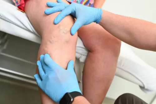
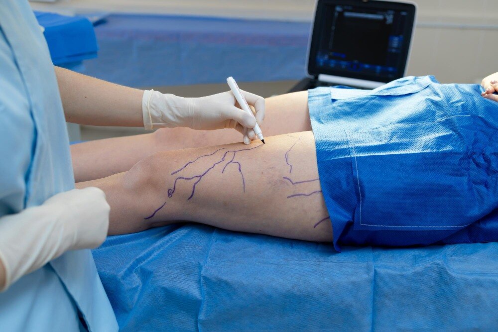
| Varicose veins or venous ulcer | Venous disease | Swelling, heaviness, skin staining; ulcer near the inner ankle |
| Deep vein thrombosis | Acute venous clot | New one-sided swelling, warmth and tenderness; seek urgent care |
| Nerve, joint or muscle pain | Back, nerve, joint or soft-tissue problem | Burning, numbness, weakness, movement-related joint pain, or pain linked to the spine |
Clinicians confirm peripheral artery disease with an ankle-brachial index (ABI). An ABI of 0.90 or less supports the diagnosis; above 1.40 can mean stiff arteries, so toe-brachial index or toe pressures help, especially with diabetes or kidney disease.
Arterial Doppler ultrasound maps flow, while CT angiography or MR angiography shows the blockage; catheter angiography is usually reserved for detailed planning or treatment. A low ABI alone does not justify angioplasty.
When is lower limb angioplasty the right treatment?
Angioplasty suitability depends on whether restoring flow will improve a meaningful problem, not on a low ankle-brachial index alone. A vascular specialist weighs the artery involved, narrowing or blockage length, degree of calcification, downstream blood flow (runoff), symptoms, walking limitation, kidney function, diabetes, previous treatment, available vein, operative risk, life expectancy and your goals.
Critical limb-threatening ischaemia—rest pain, gangrene, or a foot wound with threatened tissue—usually requires urgent revascularisation. The choice may be angioplasty, bypass, or both, depending on anatomy and surgical risk.
For intermittent claudication, treatment usually starts with:
- Supervised walking exercise
- Smoking cessation
- Antiplatelet treatment when indicated
- High-intensity cholesterol lowering
- Diabetes and blood-pressure control
Observation remains reasonable when symptoms are manageable. Consider angioplasty when walking pain still restricts work or daily activities after this programme.
| Artery or pattern | Common approach | Key consideration |
|---|---|---|
| Iliac artery blockage | Balloon angioplasty, often with stenting | Stent persistent recoil or residual narrowing |
| Femoropopliteal disease above the knee | Balloon, drug-coated balloon, selective stenting | Long, calcified segments have more restenosis |
| Below-knee or tibial disease | Balloon angioplasty | Usually reserved for tissue-threatening ischaemia |
| Long, complex disease | Surgical bypass or a combined strategy | A usable vein and surgical fitness affect durability |
Atherectomy can remove selected plaque, especially heavily calcified disease, but it is not automatically safer or more durable. Plain balloon treatment suits some short lesions; a drug-coated balloon may reduce recurrent narrowing. Medical treatment is preferred when symptoms are mild or the procedure cannot offer worthwhile benefit.
What happens before and during the procedure?
Before scheduling, your clinician reviews every medicine, including aspirin, clopidogrel, warfarin, apixaban, and diabetes tablets or insulin. Blood tests check haemoglobin, clotting status, creatinine, and estimated glomerular filtration rate (eGFR), which helps assess contrast-related kidney risk. Tell the team about any previous contrast reaction.
They may compare ankle-brachial index, toe-brachial index, arterial Doppler ultrasound, or CT angiography findings before treatment.
Fasting and medicine instructions depend on sedation, diabetes, and bleeding risk:
- Follow the stated fasting period; do not eat or drink after the instructed time.
- Do not stop warfarin, apixaban, aspirin, or clopidogrel yourself. The treating team will say whether to continue, pause, or adjust each drug.
- Ask exactly what to do with insulin, metformin, or other diabetes medicines on the procedure morning, because fasting changes glucose and kidney-safety decisions.
You lie on an X-ray table, and local anaesthetic numbs the access site. A thin catheter enters an artery, usually through the groin and sometimes the wrist. Contrast outlines the blockage on angiography; a guidewire crosses it, followed by balloon inflation to widen the channel.
A drug-coated balloon delivers medicine to reduce re-narrowing, while atherectomy removes or modifies selected heavily calcified plaque. A stent is added only when the artery recoils, significant narrowing remains, or a dissection restricts flow.
What are the risks, recovery steps, and signs of restenosis?
Post-angioplasty recovery is often shorter than recovery from bypass surgery, but it depends on the access artery, stent use, kidney function, bleeding, and whether tissue loss or infection is being treated.
Risks include bruising or bleeding at the access site, contrast-associated kidney injury, an allergic contrast reaction, arterial dissection, perforation, clot formation, distal embolisation, acute vessel closure, stent failure, and restenosis. Emergency surgery and rare limb-threatening loss of blood flow are possible.
- Follow the discharge plan for bed rest, walking limits, driving, work, and access-site care. These instructions vary with groin or wrist access, closure method, sedation, bleeding risk, and the clinician’s findings. Keep the site clean and dry as instructed; report expanding swelling or persistent bleeding.
- Take prescribed antiplatelet and cholesterol medicines exactly as directed. Stopping either independently can promote clotting, acute vessel closure, stent failure, or restenosis.
- Attend follow-up even when walking improves. Assessment can include your symptoms, foot pulses, ankle-brachial index, and duplex ultrasound; the timing depends on the treated artery and device.
- Seek prompt medical review for renewed rest pain, a cold or discoloured foot, sudden weakness, worsening pain, uncontrolled bleeding, or a wound that worsens or develops gangrene. These signs can indicate recurrent blockage or threatened tissue.
How should you compare angioplasty with bypass and long-term treatment?
Angioplasty opens the treated segment; it does not remove systemic atherosclerosis. Long-term protection still requires smoking cessation, high-intensity lipid lowering, blood-pressure and diabetes control, antiplatelet treatment when prescribed, supervised exercise therapy, foot care and wound healing support.
| Option | Main advantage | Main limitation |
|---|---|---|
| Angioplasty | Shorter recovery and less surgical trauma | Restenosis or repeat treatment, especially with long calcified lesions, poor runoff, smoking, diabetes, or kidney disease |
| Bypass | Potentially greater durability for suitable extensive disease | Larger operation, longer recovery, and dependence on a usable vein and target artery |
| Long-term treatment | Reduces cardiovascular risk and supports walking after either procedure | Does not remove an existing severe blockage or replace wound and infection treatment |
Femoropopliteal stents add a mechanical concern: bending and compression near the hip and knee can contribute to stent fracture, restenosis, or reintervention. A technically successful angioplasty is not a lifelong guarantee.
- Which artery is affected, and how long is the lesion?
- Which device is planned, and what is the chance of a stent?
- How much improvement should you expect in walking or wound healing?
- What is the repeat-treatment risk, and what are the alternatives?
- What happens if the wire cannot cross the blockage?
Arrange transport after sedation, attend planned imaging appointments, inspect wounds as instructed, take medicines consistently, and keep cardiovascular follow-up. Manage infection or tissue loss urgently. In Thane, Radiance Clinic can provide an assessment combining interventional radiology and vascular medicine to discuss these choices.
Related service
Vascular And Interventional Radiology Peripheral vascular disease (PVD) is a condition, in which a build-up of fatty deposits in the arteries which reduces the inner diameter of the... View service → |
Frequently asked questions
Which leg symptoms point to an artery problem?
Calf or thigh pain during walking that improves with rest suggests reduced arterial flow. Rest pain, a cold or discoloured foot, and nonhealing wounds indicate more severe disease.
When is lower limb angioplasty the right treatment?
Angioplasty is considered when an artery narrowing causes disabling walking pain, threatens the limb, or needs revascularisation and the anatomy is suitable for catheter treatment.
What happens before and during lower limb angioplasty?
Assessment includes symptom history, pulse examination, ankle-brachial pressure testing, and imaging such as duplex ultrasound or CT angiography. During the procedure, a catheter delivers a balloon to widen the narrowing; a stent can support the artery when required.
What are the risks and recovery steps after angioplasty?
Risks include bleeding, bruising, artery injury, clot formation, kidney effects from contrast, infection, and re-narrowing. Follow wound-care, walking, medication, and follow-up instructions, and seek urgent help for worsening pain, a cold or pale foot, heavy bleeding, or sudden weakness.
How does angioplasty compare with bypass and long-term treatment?
Angioplasty usually involves a catheter-based recovery, while bypass uses a graft to route blood around a blockage and requires surgery. Both still require walking therapy, prescribed antiplatelet treatment, cholesterol and blood-pressure control, diabetes management, and smoking cessation.